Clearly present the unique features of your patient’s condition
with the aide of these elegant anatomical illustrations.




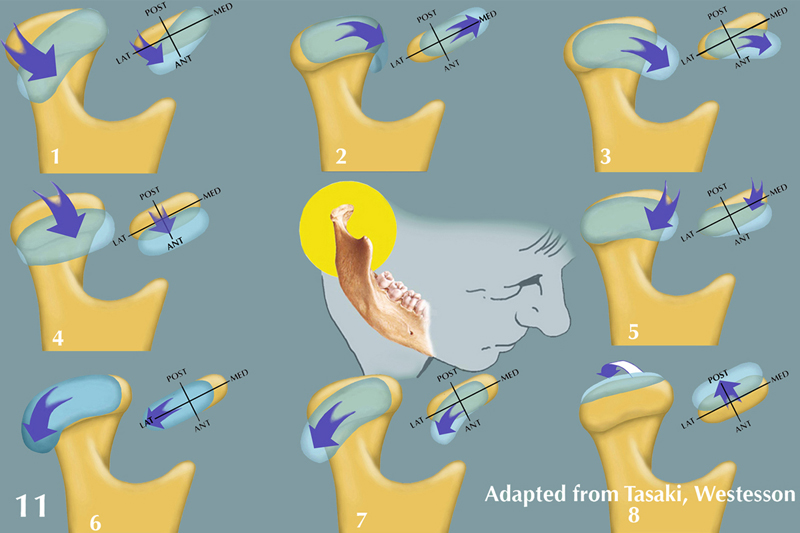
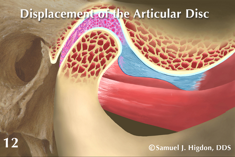
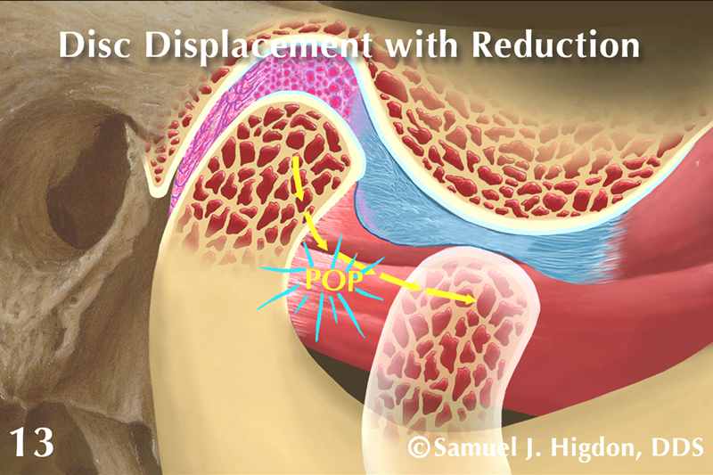
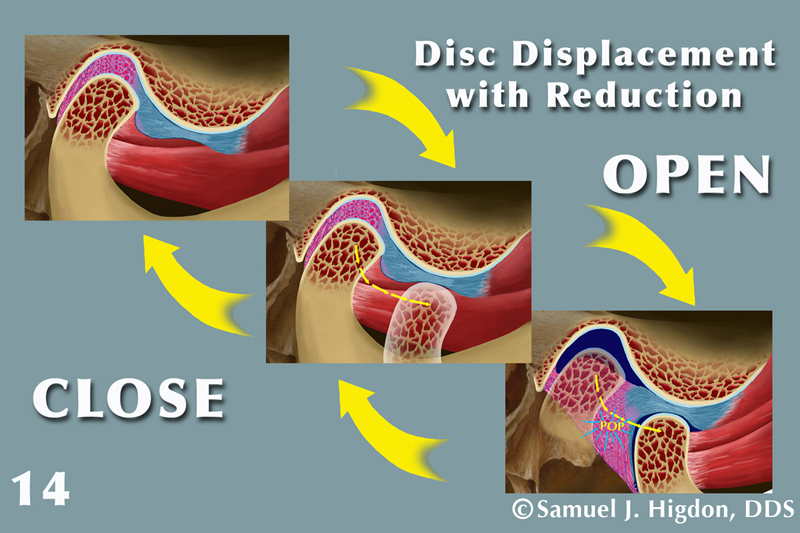
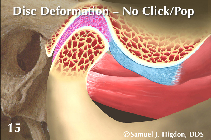

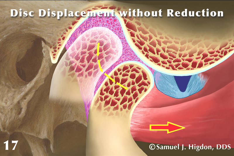



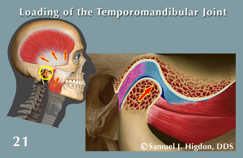
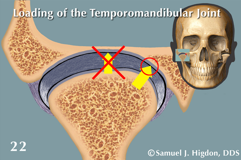
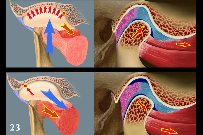

Accompanying the Patient Education Aid illustrations is also extensive instructional text from which you will learn the most important anatomical and functional information about these often-confusing disorders.
“Illustrated Anatomy of the Temporomandibular Joint in Function / Dysfunction” is available in two sizes.
These are intended to be a patient education aide. The illustrations in the album allow the dentist or other health care provider to explain to their patient the condition of the TM joint that relates to their signs and symptoms. In addition to the illustrations is an extensive text that is provided to inform the dentist or other health care provider with an in-depth description of what is currently known about TMJ anatomy and function.
Small album
The small album (4” x 6”) contains 23 illustrations. It is accompanied by a booklet that provides the text described above, together with thumbnail illustrations that correspond with the illustrations in the album, plus several other illustrations that will help clarify the text.
Large album
The large album (9.5” x 11.5”) contains the same but much larger illustrations and also contains the text, together with the thumbnail illustrations.
Links to other sites
About
Buy Now


